As a further step of our comparative study of Pisum seed proteins (1,
2, 3), we report now on an electrophoretic analysis of urea-treated legumin
fraction from various Pisum ecotypes. The analyzed material comprised 21
Pisum lines originating from the Weibullsholrn collection, the John Innes
Institute, and from other sources. Isolation of legumin fraction from total
globulin extracts was performed by isoelectric precipitation at pH 4.7. Poly-
acrylamide gel electrophoresis (PAGE) of urea-treated legumin was conducted
in anodic and cathodic buffer systems to achieve resolution of acidic and basic
components, respectively.
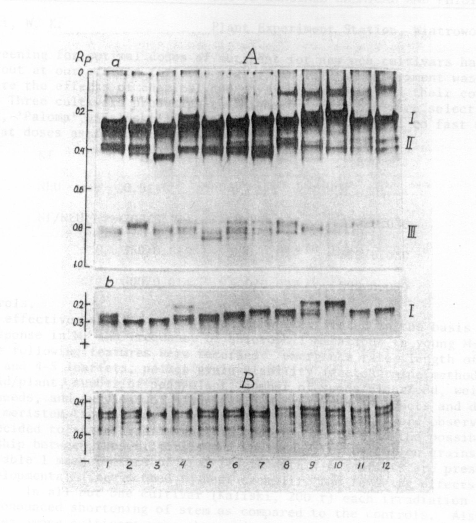
Electrophoretic banding patterns obtained in an anodic buffer system show
3 variant zones, I-III (Fig. lA-a); slow-moving bands of zone I are the major
bands which are better resolved when smaller amounts of protein are subjected
to electrophoresis (Fig. lA-b). Variation within each of the zones is shown
for 12 Pisum lines. However, the overall variation is not entirely revealed
since in individual lines electrophoretic banding patterns of particular zones
form specific combinations. Within the investigated material 17 distinct
patterns of anodic bands were observed.
Urea gels obtained in a cathodic buffer system revealed two, three, or
four bands of basic proteins in particular lines (Fig. IB). The investigated
lines showed 6 distinct patterns of cathodic bands.
In agreement with the finding of Thomson et al. (4), urea-gel electro-
phoresis proved to be very useful in analysis of Pisum legumin. The previously
reported SDS-gel electrophoretic patterns of legumin fractions from distant
Pisum lines (3) showed P. fulvum as a distinct taxon and differences between
P. fulvum lines. Urea-gel electrophoretic patterns, presented here, confirmed
the above data and, in addition, revealed marked variation within lines classi-
fied as P. elatius, P_. humile, and P. sativum. Moreover, P. abyssinicum
proved to have a "species-specific" legumin pattern.
1. Hurich, J., Parzysz, 11., Przybylska, J. 1977. Genetica Polonica
18:241-252.
2. Przybylska, J., Blixt, S., Hurich, J., Zimniak-Przybylska, Z. 1977.
Genetica Polonica 18:27-38.
3. Przybylska, J., Hurich, J., Zimniak-Przybylska, Z. 1978. PNL 10:66-G7.
4. Thomson, J. A., Schroeder, H. E., Dudman, W. F. 1978. Austr. J. Plant
Physiol. 5:263-280.
1/ - This work was performed under Government Project PR-4.